Monday, July 22, 2013

Concentração sérica de insulina em cães apresentando hipoadrenocorticismo / Insulin concentration in dogs with hypoadrenocorticism


Insulin concentrations in dogs with hypoadrenocorticism

  • a Royal (Dick) School of Veterinary Studies, The Roslin Institute, Division of Clinical Sciences, The University of Edinburgh, Hospital for Small Animals, Easter Bush Veterinary Centre, Roslin, Midlothian EH25 9RG, UK
  • b Division of Small Animal Clinical Sciences, School of Veterinary Medicine, College of Medical, Veterinary and Life Sciences, University of Glasgow, 464 Bearsden Road, Glasgow G61 1QH, UK
  • c Cambridge Specialist Laboratory Services Ltd., P.O. Box 967, Stapleford, Cambridge CB2 5XY, UK

    Abstract

    Hypoglycaemia is frequently identified in canine cases of hypoadrenocorticism. Potassium and glucose cellular uptake are intimately linked by insulin. We hypothesized that in canine hypoadrenocorticism, hyperkalaemia would stimulate insulin release as a protective mechanism, translocating potassium from the extracellular compartment to the intracellular compartment and also lower glucose concentrations. Serum insulin concentrations were measured in 11 consecutive cases of canine hypoadrenocorticism which were hyperkalaemic and 33 dogs with non-adrenal illness. There was no significant difference between insulin concentrations in the two populations, and no correlation between insulin and potassium concentration in the hypoadrenal group. Thus, no support for the hypothesis was found, although multiple other factors such as pH and osmolality may be obscuring an effect.

    Keywords

    • Canine
    • Insulin
    • Potassium
    • Hyperkalaemia
    • Hypoadrenocorticism
  • Signalment
    Glucose/mmol/L
    Potassium/mmol/L
    Insulin/μIU/mL (ref. range 4.4-17.6)
    English springer spaniel, 5years 5month, IM[low *]
    3.8
    6.3a
    3.8b
    Labrador 5years 3month, NF[low *]
    5.4
    6.8a
    2.6b
    Labradoodle 1years 1month IM[low *]
    4.0
    7.7a
    7.1
    Collie-cross, 1years 8month, NF[dagger]
    4.8
    6.85a
    33a
    St. Bernard, 1years 9month, IM[low *]
    5.1a
    6.4a
    8.5
    Chihuahua, 7years 2month, NF[low *]
    4.9
    8.8a
    <2.0b
    Standard poodle, 2years, IF[dagger]
    7.4a
    6.24a
    2.9b
    Cocker spaniel, 7years, NF[dagger]
    4.67
    10a
    27a
    German short-haired pointer, 6years, NM[dagger]
    2.9b
    8a
    <2.0b
    Border collie, 6years, NM[low *]
    4.7
    7.8a
    8.6
    English springer spaniel, 2years 6month, IF[low *]
    4.9
    6.9a
    3.5b
    (b) Below reference range.

Wednesday, July 17, 2013

Acuracia diagnostica do teste rivalta para peritonite infecciosa felina / Diagnostic accuracy of the Rivalta test for feline infectious peritonitis

Diagnostic accuracy of the Rivalta test for feline infectious peritonitis

  1. Yvonne Fischer1
  2. Carola Sauter-Louis2,
  3. Katrin Hartmann1,*
Article first published online: 22 AUG 2012
DOI: 10.1111/j.1939-165X.2012.00464.x
Veterinary Clinical Pathology

Veterinary Clinical Pathology

Volume 41Issue 4pages 558–567December 2012

Keywords:

  • Cat;
  • diagnosis;
  • effusion;
  •  FIP ;
  • sensitivity;
  • specificity

Background

The Rivalta test has been used routinely in Europe to diagnose feline infectious peritonitis (FIP) in cats with effusions, but its diagnostic accuracy is uncertain.

Objectives

The objectives of this study were to calculate sensitivity, specificity, and positive (PPV) and negative (NPV) predictive values of the Rivalta test for FIP and to identify correlations between a positive Rivalta test and variables measured in effusion fluid and peripheral blood.

Methods

In this retrospective study, medical records of cats with effusions were reviewed, and cats with conclusive results for the Rivalta test were included. The prevalence of FIP in this population was determined, and sensitivity, specificity, and PPV and NPV of the Rivalta test were calculated. Variables measured in effusion fluid and peripheral blood were compared between cats that had positive or negative Rivalta tests using the Mann–Whitney U-test and multivariate analysis.

Results

Of 851 cats with effusions, 782 had conclusively positive or negative results for the Rivalta test. A definitive final diagnosis was made in 497 of these cats. Prevalence of FIP in cats with effusion and a conclusive Rivalta test result was 34.6%. The Rivalta test had a sensitivity of 91.3%, specificity of 65.5%, PPV of 58.4%, and NPV of 93.4% for the diagnosis of FIP. These values increased when cats with lymphoma or bacterial infections were excluded, or when only cats ≤ 2 years were considered. Increased effusion cholesterol concentration and specific gravity as well as decreased serum albumin:globulin ratio and hyperbilirubinemia were positively correlated with positive Rivalta test results.

Conclusions

Sensitivity, specificity, and PPV of the Rivalta test for the diagnosis of FIP were lower than previously reported except when used in young cats. The components in effusions that lead to a positive Rivalta test remain unknown, but the positivity is not simply related to high total protein concentration.


Saturday, July 13, 2013

Intoxicação por ivermectina em 17 collies / Ivermectin toxicity in 17 collies

Ivermectin Toxicity in 17 Collies

  1. Kate Hopper1,*
  2. Janet Aldrich1
  3. Steve C. Haskins2
Article first published online: 28 JUN 2008
DOI: 10.1111/j.1939-1676.2002.tb01611.x
Journal of Veterinary Internal Medicine

Journal of Veterinary Internal Medicine

Volume 16Issue 1pages 89–94January 2002

vermectin is widely used in veterinary medicine as an anthelminthic and generally has a wide margin of safety, but Collies are prone to ivermectin toxicity. Two groups of Collies were presented to the University of California Veterinary Medical Teaching Hospital (VMTH) with ivermectin toxicity. The medical records of the 2 groups of Collies were reviewed retrospectively. Group I comprised 5 adult Collies that received at least 400 μg/kg ivermectin PO and were presented to the VMTH 3 hours after intoxication. These Collies showed marked clinical signs on presentation. Three of these dogs required mechanical ventilation and were euthanized for financial reasons; the remaining 2 dogs were comatose but recovered in 5–7 days. Group II was comprised of 12 adult Collies presented to the VMTH 2 days (n = 10) and 5 days (n = 2) after subcutaneous injection of 200–250 μg/kg ivermectin. These animals showed greater variation in severity of illness among individuals; 5 animals progressed to stupor or coma, whereas 4 animals remained ambulatory. Most of these dogs' clinical signs deteriorated from the day of intoxication until approximately day 6, from which time they showed gradual but steady improvement. All of the Collies in this group survived, but it took 3 weeks for most of them to recover. Collies suffering from ivermectin toxicity can have a severe and prolonged clinical course requiring intensive nursing care. Respiratory, cardiovascular, and nutritional support may all be required. With appropriate care, however, the prognosis for complete recovery is good.


Tuesday, July 9, 2013

Trombose de veia porta em 33 cães / Portal vein thrombosis in 33 dogs

You have full text access to this content

Portal Vein Thrombosis in 33 Dogs: 1998–2011

  1. M. Respess1,*
  2. T.E. O'Toole1
  3. O. Taeymans1
  4. C.L. Rogers2
  5. A. Johnston1,
  6. C.R.L. Webster1
Article first published online: 28 FEB 2012
DOI: 10.1111/j.1939-1676.2012.00893.x
Journal of Veterinary Internal Medicine

Journal of Veterinary Internal Medicine

Volume 26Issue 2pages 230–237March-April 2012

Keywords:

  • Coagulation;
  • Coagulopathy;
  • Liver disease;
  • Thromboembolism

Background

Portal vein thrombosis (PVT) has been reported infrequently in dogs.

Objectives

To characterize the presentation, associated disease conditions, and outcome in dogs with PVT.

Animals

Client-owned dogs with a diagnosis of PVT and a complete medical record.

Methods

Records were retrospectively analyzed for presentation, history, physical examination, clinicopathologic data, diagnostic imaging, treatment, and outcome.

Results

Thirty-three dogs were included. Common clinical signs were vomiting, diarrhea, abdominal pain, ascites, and signs of hypovolemic shock. Associated disease conditions included hepatic (14/33), neoplastic (7/33), immune (5/33), and infectious (4/33) diseases, protein-losing nephropathy (3/33), hyperadrenocorticism (2/33), protein-losing enteropathy (1/33), and pancreatitis (1/33). Fourteen dogs were receiving glucocorticoids at the time of diagnosis. Twenty-nine dogs had at least 1 predisposing condition for venous thrombosis, and 11 had 2 or more. Thrombocytopenia (24/33), increased liver enzyme activity (23/33), and hypoalbuminemia (20/33) were common laboratory abnormalities. Clinical syndromes at the time of PVT diagnosis included shock (16/33), systemic inflammatory response syndrome (SIRS), (13/33) and disseminated intravascular coagulation (3/33). Twenty-four dogs had acute and 9 had chronic PVT. Multiple thrombi were found in 17/33 dogs. Nineteen dogs survived to discharge. Dogs treated with anticoagulant therapy were more likely, whereas those with acute PVT, multiple thromboses or SIRS were less likely to survive.

Conclusions and Clinical Importance

Hepatic disease is a common pre-existing condition in dogs with PVT. PVT should be considered in dogs with risk factors for venous thrombosis presenting with abdominal pain, ascites, and thrombocytopenia. Studies evaluating anticoagulant therapy in the management of PVT are warranted.

Friday, July 5, 2013

Tratamento da anemia hemolítica imunomediada com ajuste individual da dosagem de heparina / Treatment of Immune-Mediated Hemolytic Anemia with Individually Adjusted Heparin Dosing in Dogs

Treatment of Immune-Mediated Hemolytic Anemia with Individually Adjusted Heparin Dosing in Dogs

  1. S.E. Helmond1
  2. D.J. Polzin1
  3. P.J. Armstrong1
  4. M. Finke1
  5. S.A. Smith2
Article first published online: 6 APR 2010
DOI: 10.1111/j.1939-1676.2010.0505.x
Journal of Veterinary Internal Medicine

Journal of Veterinary Internal Medicine

Volume 24Issue 3pages 597–605May/June 2010


Keywords:

  • Anti-Xa;
  • Survival;
  • Thromboembolism;
  • Thrombosis
Background: A major cause of death in dogs with immune-mediated hemolytic anemia (IMHA) is thromboembolism. Previous studies suggest unfractionated heparin (UH) is not effective in preventing thromboembolism in IMHA; however, subtherapeutic dosing could explain the seeming lack of efficacy.
Hypothesis: Providing therapeutic plasma concentration of UH by individually adjusting doses based on antifactor Xa activity would improve survival in IMHA.
Animals: Fifteen dogs with primary IMHA.
Methods: Randomized, prospective, controlled clinical trial. Dogs received standardized therapy for IMHA and either constant dose (CD) (150 U/kg SC) (n = 7) or individually adjusted dose (IAD) (n = 8) UH, monitored via an anti-Xa chromogenic assay, adjusted according to a nomogram. UH was administered every 6 hours until day 7, and every 8 hours thereafter. UH dose was adjusted daily in IAD dogs until day 7, weekly until day 28, then tapered over 1 week. Dogs were monitored for 180 days.
Results: At day 180, 7 dogs in the IAD group and 1 in the CD group were alive (P= .01). Median survival time for the IAD group was >180 days, and 68 days for the CD group. Thromboembolic events occurred in 5 dogs in the CD group and 2 dogs in the IAD group. Doses of UH between 150 and 566 U/kg achieved therapeutic anti-Xa activity (0.35–0.7 U/mL).
Conclusions and Clinical Importance: This study suggests that IAD UH therapy using anti-Xa monitoring reduced case fatality rate in dogs with IMHA when compared with dogs receiving fixed low dose UH therapy.


Tuesday, July 2, 2013

Avaliação clínica e manejo do hemoperitoneo em cães / Clinical evaluation and management of hemoperitoneum in dogs

Clinical evaluation and management of hemoperitoneum in dogs

  1. Lee V. Herold DVM, DACVECC1
  2. Jennifer J. Devey DVM, DACVECC2
  3. Rebecca Kirby DVM, DACVIM, DACVECC3
  4. Elke Rudloff DVM, DACVECC3
Article first published online: 4 FEB 2008
DOI: 10.1111/j.1476-4431.2007.00265.x
Journal of Veterinary Emergency and Critical Care

Journal of Veterinary Emergency and Critical Care

Volume 18Issue 1pages 40–53February 2008


Keywords:

  • damage-control surgery;
  • hemorrhagic shock;
  • hemostasis;
  • intra-abdominal hemorrhage;
  • trauma

Abstract

Objective: Review the clinical presentation, assessment, resuscitation, and medical and surgical management of dogs with hemoperitoneum.
Etiology: Hemoperitoneum is defined as free intra-abdominal hemorrhage. Hemoperitoneum occurs from traumatic and nontraumatic causes. Common etiologies include atraumatic rupture of intra-abdominal masses, coagulopathies, as well as blunt, and penetrating trauma to the abdomen.
Diagnosis: Definitive diagnosis of hemoperitoneum entails demonstration of free intra-abdominal blood via paracentesis or diagnostic peritoneal lavage. Imaging and other diagnostic tests including coagulation studies may help to determine underlying causes of hemoperitoneum or concurrent organ dysfunction.
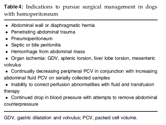
Therapy: Goals of therapy for patients with hemoperitoneum include maintenance and restoration of effective circulating volume, maintenance and restoration of oxygen-carrying capacity, and arrest of hemorrhage. These goals can be achieved via fluid resuscitation, administration of blood products or hemoglobin-based oxygen carriers, as well as application of abdominal counterpressure, and surgical intervention. Surgery usually is required for bleeding intra-abdominal neoplasms. Emergency surgery is recommended for hemorrhaging patients with penetrating trauma, gastric dilatation and volvulus, bleeding cysts, liver lobe torsion, splenic torsion, and any other condition resulting in organ ischemia.
Prognosis: Prognosis in patients with hemoperitoneum may depend on the underlying cause and concurrent injuries.


Monday, July 1, 2013

Manejo da ruptura uretral traumática em 11 gatos usando o alinhamento primário com sonda uretral / Management of traumatic urethral rupture in 11 cats using primary alignment with a urethral catheter

Veterinary and Comparative Orthopaedics and Traumatology (VCOT)
2008: Issue 1 2008

Management of traumatic urethral rupture in 11 cats using primary alignment with a urethral catheter


Management of traumatic urethral rupture in 11 cats using primary alignment with a urethral catheter

F. Meige1, S. Sarrau2, A. Autefage3
1Clinique Vétérinaire Occitanie, Toulouse, France 2Clinique Vétérinaire de Parme, Biarritz, France 3National Veterinary School of Toulouse, Department of Small Animal Clinical Sciences, Toulouse cedex 03, France

Summary

Objective: To assess the management of traumatic urethral ruptures using a systematic attempt at urethral catheterisation. Methods: Eleven cats that had been diagnosed with a traumatic urethral rupture over a fiveyear period were included in this study. Rupture was assessed by positive-contrast retrograde urethrography. An attempt was made to manage these ruptures by placing an indwelling urethral catheter. Results: The placement of an indwelling urethral catheter, could be performed in 10 out of 11 cats, which indicated a partial rupture. In one cat, catheterisation was not possible because of complete urethral rupture. A urethral catheter was placed into the urinary bladder via the urethra in five animals. In the other five animals, urethral catheterization required cystotomy using a modified ‘inside to outside’ technique. Urethral catheter removal occurred between five and 14 days after placement. Urethral healing was accomplished in all cases of catheterisation with a good outcome in eight of the 10 cats. Two cats showed clinical signs related to stricture formation during the follow-up period (seven to 72 months). Retrograde urethrography provided a definitive diagnosis of partial rupture in only six of the 10 cases of partial rupture. Clinical significance: Results suggested that primary alignment with the placement of a urethral catheter should be attempted for the management of traumatic urethral ruptures in cats. Gentle urethral catheterisation seems to be an effective procedure for diagnosing and dealing with partial urethral rupture in cats. Clinical stricture formation can occur, but the risk does not seem to be high. Further studies are warranted in order to define the ideal duration for maintenance of an indwelling urethral catheter in cases of traumatic partial urethral rupture.

Keywords

cats, Trauma, rupture, Urethra, catheterisation